From coast to coast and around the world, scientists like Andrew
F. Leuchter, M.D., and Michael Levine, Ph.D., are engaged in the quest for
Huntington’s disease treatments.
During May, Huntington’s Disease Awareness Month, I want to call
attention to the critical work of Drs. Leuchter and Levine on the West Coast. They exemplify the
partnership of scientists and physicians with the HD community, aiming to
advance potential remedies into crucial clinical trials.
Drs. Leuchter and
Levine, faculty researchers at the renowned Semel Institute for Neuroscience and Behavior at the University of California, Los Angeles
(UCLA), are collaborating on a project that could ultimately lead to new drugs.
In the near term, they aim to understand more fully the electrical signals that
naturally but abnormally emanate from the brains of HD patients and
presymptomatic carriers of the HD gene mutation like me.
“Most of the brain’s energy goes to creating electrical gradients
– electrical impulses – but we haven’t been very good at using that for
diagnosis and treatment,” Dr. Leuchter said during a March 20 interview in his
office at the Semel Institute. He and Dr. Levine aim to “decipher the signals
that are coming out of the brain.”

The Semel Institute for Neuroscience and Behavior (photo by Gene Veritas)

The Semel Institute for Neuroscience and Behavior (photo by Gene Veritas)
Measuring brain energy
A psychiatrist specializing in depression and Alzheimer’s
disease, Dr. Leuchter (pronounced LUKE-ter) frequently employs quantitative
electroencephalography (quantitative EEG) to measure the energy emitting from
people’s brains. One example: a group of 27 HD subjects he and others observed
for a study published in 2010 and funded by CHDI Foundation, Inc., the nonprofit virtual biotech
dedicated exclusively to the discovery of HD treatments.
Allan Tobin, Ph.D., at the time the head of UCLA’s Brain Research
Institute and a senior scientific advisor at CHDI, had asked colleague Leuchter
for assistance in finding HD biomarkers, signals that reveal the progression of the disease and/or the
effectiveness of a medication.
As the number of HD clinical trials expands exponentially, the search for useful biomarkers has become one of the hottest areas in Huntington's disease research. (Click here to read about one new potential biomarker.)
As the number of HD clinical trials expands exponentially, the search for useful biomarkers has become one of the hottest areas in Huntington's disease research. (Click here to read about one new potential biomarker.)
As Dr. Leuchter pointed out, neurological and psychiatric
disorders are “much more limited in diagnostic tests for the organ that we are
studying than any other branch of medicine.” Cardiologists insert catheters
into the heart, and gastroenterologists use scopes to view the stomach and
intestines.
“If you’re a psychiatrist, we talk to people, which is great, but
we don’t have physiologic tests that guide decision-making,” he added.
Scientists and doctors rarely put electrodes in living human
brains or take biopsies of brain tissue. However, they have been measuring
brain energy with EEGs for more than a century, Dr. Leuchter explained.
As he demonstrated in his lab (see photo below), today patients
undergoing testing wear a cap with 35 separate EEG electrodes, or contacts,
that touch the head. The attending researcher stretches the cap over the
patient’s head. In contrast with the traditional EEG, which involves one-by-one
placement of the electrodes on the head, this method is quick, efficient, and
less burdensome to patients, he noted.
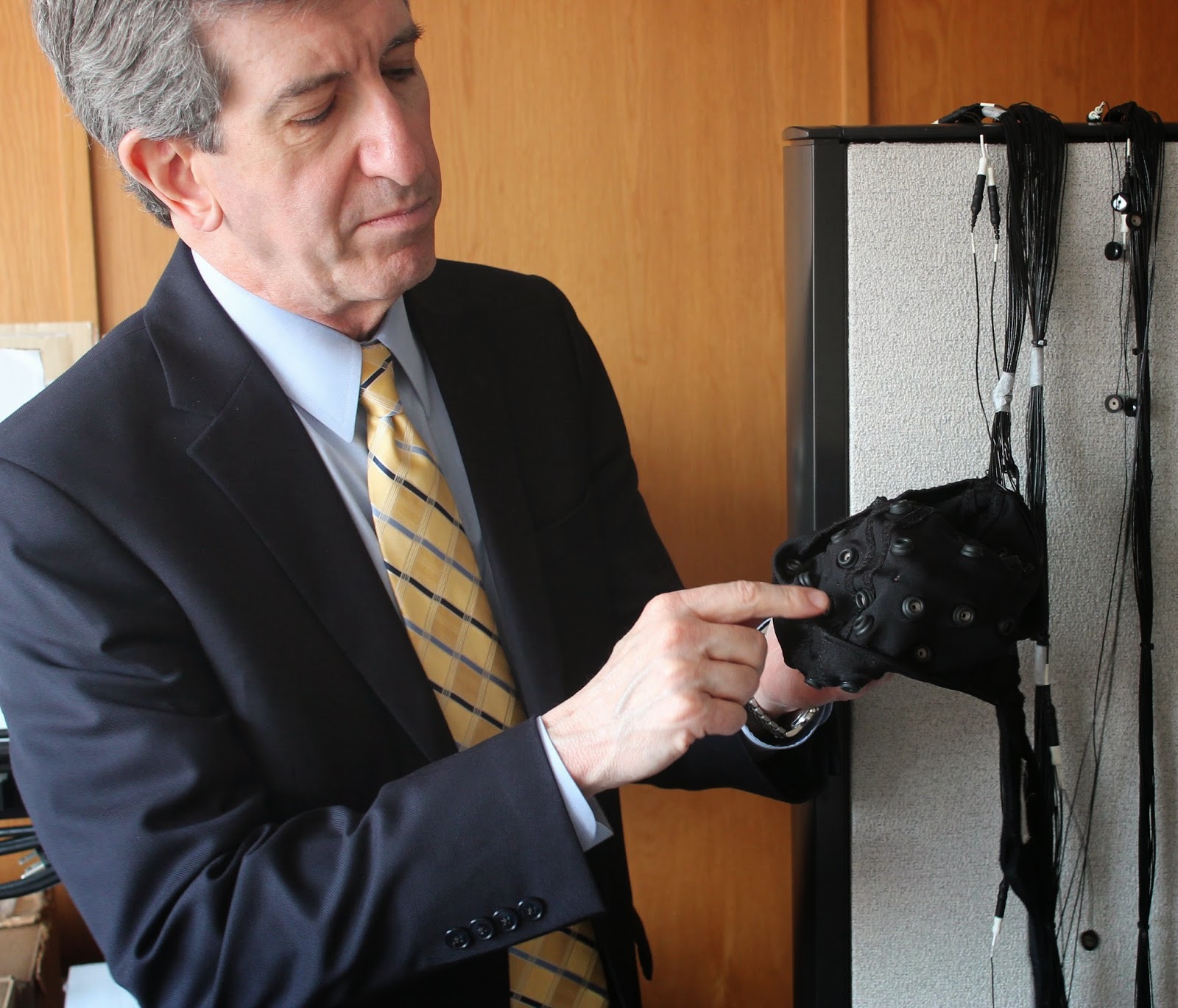
Above, Dr. Andrew Leuchter points out the electrodes on the EEG cap worn by research subjects. Below, he explains digitized EEG readings displayed on a computer monitor. (photos by Gene Veritas)

“We find that this helps to standardize our measurements of brain
activity, and that we can place the electrodes in about 15 minutes,” Dr. Leuchter said.
EEG is inexpensive, convenient, and easy to administer.
Additionally, it does not expose patients to radiation or require them to lie
inside a machine such as an MRI scanner, he noted.
“You can tote it wherever you like,” he said of the EEG device.
The brain’s pacemaker
As they had hoped, Dr. Leuchter and three other UCLA researchers
discovered abnormal EEG readings in HD patients with just mild symptoms.
“But the really intriguing thing there was that, even in people
who were gene-positive but premanifest, we could see differences in brain
function estimated 15, 20 years out from diagnosis,” Dr. Leuchter said, referring to signals of
future decline. “So we thought this
could be something that could be useful for treatment development.”
As Dr. Leuchter explained, “the brain like the heart has pacemakers.”
Healthy brains produce lots of high-frequency waves. Brain illnesses commonly
result from changes in the firing of the pacemaker, resulting in a greater
quantity of low-frequency waves.
“What we found is that years before people start to show symptoms
with Huntington’s, they’re producing more low-wave energy,” Dr. Leuchter said. “So it’s a very subtle indicator that the
pacemaker of the brain is starting to slow down.”
Scientists cannot predict the actual onset and
progression of symptoms from EEG signals. However, as noted below, they did discover a correlation between the
severity of genetic mutation and EEG readings.
Clear genetic impact on the brain
Furthermore, the team observed that, in contrast with healthy
brains, the distribution of different types of waves across the different
regions of the HD brains became more uniform. “The regions of the brain start
to look more similar than different,” he explained.
Researchers have not yet discovered what this phenomenon means.
“We know that the brain has enormous functional reserve and that
people call on every cognitive and emotional resource they’ve got to try to
keep everything functioning at optimal efficiency,” Dr. Leuchter continued. “I don’t think we know what’s compensatory
and what’s an early sign of illness.”
Reflecting on another facet of the research, Dr. Leuchter
explained that, in general, brain function tests do not correlate with genetic
factors.
However, he and his team did find a correlation between
the degree of HD genetic mutation and the severity of the changes in the EEG
readings.
“Nobody had seen that,” he recalled. “We got excited about that,
and that’s what we’ve been trying to follow up on.” These findings will
contribute to the search for biomarkers and treatments, as explained below.
Examining brain tissue
A neurophysiologist and veteran basal ganglia researcher, since the late
1960s Dr. Levine has studied these deep, inner
parts of the brain that control such actions as voluntary movements. He began to study HD in the 1990s as genetic mouse models with HD-like symptoms became available. His lab has published more than two dozen papers about these mice.
The nuclei of the basal ganglia are significantly compromised in HD, especially in the striatum. Specifically, Dr. Levine has examined how neurons communicate with each other in the cortex and striatum at cellular and molecular levels using tissue from the HD mouse models.
The nuclei of the basal ganglia are significantly compromised in HD, especially in the striatum. Specifically, Dr. Levine has examined how neurons communicate with each other in the cortex and striatum at cellular and molecular levels using tissue from the HD mouse models.
One of the latest techniques for studying the cells in the HD mouse models is
optogenetics, in which specific types of brain cells are stimulated with
light.
“I can look very closely at mechanisms,” Dr. Levine explained. “I
know which types of neurons I am looking at and how they change at a very
mechanistic level.”

Michael Levine, Ph.D., veteran HD researcher (photo by Gene Veritas)
Two key goals
Melding approaches, and with the expectation of CHDI support,
Drs. Leuchter and Levine now seek to answer two important questions.
The first involves comparing EEG data from both mice and humans
to refine the search for biomarkers. Researchers have already made the key
discovery of EEG signals common to mice and humans.
“It’s actually pretty uncommon in science that you can see a very
similar signal across species, that you can see something very similar in the
brains of humans and the brains of animals,” Dr. Leuchter said.
If the Leuchter-Levine project confirms the degree of that
similarity, that could mean potential
drugs tested in mice could ultimately be used for human clinical trials, Dr. Leuchter observed.
The second question focuses on the testing in HD mice of a
CHDI-developed compound aimed at lowering the amount of mutant huntingtin
protein, the major culprit in the disease.
“If we do see a link between lowering of mutant huntingtin and change
in the EEG biomarker, this could be used to develop a number of therapeutic
agents,” Dr. Leuchter said. “A
whole line of research could develop out of this.”
From molecule to the whole brain
Drs. Leuchter and Levine estimated the project will take two
years to complete.
As Dr. Levine put it, researchers hope the CHDI-developed
compound will restore the EEG signals in HD patients to normal.
Dr. Leuchter reflected on the significance of the project and his
collaboration with Dr. Levine: “The fact that in something like Huntington’s
disease you’ve got a protein that is affecting how the nerve cells are
functioning and altering the way they produce and utilize energy – it’s really
a gateway to understanding the connection between what is going on at the
deepest molecular level of the cell and what we’re able to see with the brain
waves the individual is putting out. We can actually potentially link
everything going from the level of the gene all the way to whole-brain
function.”
In another potential future project, Dr. Leuchter would like to
obtain EEG readings from asymptomatic gene carriers over two to three years to
better measure the changes in signals over time.

Drs. Leuchter and Levine (photo by Gene Veritas)
Participation and a positive attitude
Both researchers expressed gratitude to the HD community and
fellow HD researchers for their dedication to the cause.
“There are not that many people with this illness, so people get
asked a lot to participate in different studies where they’re poked or prodded
or scanned,” Dr. Leuchter said. “We
are very grateful to those who are so generous with their time, because without
their help we could not conduct these research studies.”
Dr. Levine added that he is impressed with the “very positive
and sharing attitude of the investigators who do research in HD and who are
looking to help the patients.”
While interviewing these two researchers, as an individual racing
against the genetic clock of HD, I was once again moved to witness the
creativity and enthusiasm of scientists engaged in the quest to save affected families from the devastation of Huntington’s.
1 comment:
I am praying for these doctor's success.
Post a Comment